MATSUI Group


Explore the mystery of electron spins!

menu

Photoelectron Momentum Microscope for μm-material electronic and spin structure visualization

The importance of characterizing electronic properties of μm- and nm-scale world cannot be overemphasized. Understanding based on the atomic level characterization methods is becoming increasingly important in elucidating new quantum phenomena and developing functional materials and devices. In photoelectron spectroscopy (PES), the photoelectrons emitted from the sample surface irradiated with X-rays are measured to clarify the composition and electronic structure of the sample. So far, high-resolution electron analyzers have been developed to measure the valence band dispersion of crystalline samples, but they average information over a wide field of view. However, the most interesting materials and useful devices are often micrometer-scale polycrystalline or highly integrated structures. Therefore, a high-specification electron energy and momentum analysis apparatus that also has microscope functions was desired. The momentum microscope, a combination of projection-type electron analyzer and photoelectron microscope, simultaneously realizes a microscope function for magnifying and observing minute parts of complicated-structured samples with element selectivity and a spectroscopy function for visualizing electron behavior (momentum) that determines the electronic properties of a sample. Now we have introduced the latest momentum microscope at the undulator-based soft X-ray beamline of UVSOR providing the optimal energy light for valence bands and core levels of various elements. Microscopy with a spatial resolution of 50 nm and photoelectron spectroscopy (field of view: 2 μm) with energy / momentum resolutions of 20 meV / 0.01 Å-1 at 9 K (-264 ºC) are successfully demonstrated. In addition to the features of momentum microscope, the sample temperature can be freely changed up to 400 K (127 ºC), which enables in-situ observation of phase transition in the materials. One can investigate how the composition, structure, and electronic state of materials are related to electronic properties and functions. A method to directly observe the electronic structure of μm-sized materials are realized. The momentum microscope opens the door to direct observation of the Fermi surface and band structure of μm-sized targets such as surface atomic sites, thin films and interfaces, molecular adsorbates, and polycrystals, which was difficult with conventional electron energy analyzers. It enables electronic property and function analysis from a new point of view, such as direct observation of structure and electronic state changes where superconductivity and catalytic activity are occurring. In addition to contributing to basic science and applied research, we aim to make this new pioneering analyzer and analysis method a precursor to measurement technology innovation.
物質中では電子は原子に散乱され、互いに干渉しあいながら、波のように運動しています。光を照射し、電子を真空中に取り出して計測する光電子分光法は、こうした物質中の電子の素性を原子レベルで解明する有力な手法として発展してきました。面白いことに、電子は波の性質を保持したまま光電子として真空中に飛び出すため、光電子の角度分布には、物質中での電子の運動や原子の並び方に由来する実に美しい波紋文様(光電子ホログラム)が現れます。光電子が織りなす「アート」を物理法則に基づいて解析すると、時として原子の世界と実用的な技術が結び付く発見に至る、それが私たちの研究の醍醐味です。 私たちは分子研極端紫外光研究施設(UVSOR)に世界最先端の光電子運動量顕微鏡の実験拠点を新設しました。光電子運動量顕微鏡は、光電子ホログラムを可視化する新しい発想の分析器で、物性が発現している微小領域に照準を合わせ、実際にその物性を司る電子状態にズームインすることができます。私たちが特に注目する電子スピンは特異な磁気特性・電気伝導・光熱励起・化学反応など様々な量子現象の源泉です。そこで、私たちは独自の3Dスピンベクトルイメージング装置や元素選択的な共鳴光電子回折分光法を創案しました。これらを光電子運動量顕微鏡と融合し、電子物性・機能が発現する現場を直接分析する研究を進めています。独自の分析装置・分光手法を駆使して電子の運動を精緻・包括的に解析し、まだ誰も知らない新奇現象を捕まえ、電子・スピン物性科学の新展開を目指します。

Highlights

カゴメ格子超伝導を担う電子軌道を解明 東北大―佐藤・中山グループと共同研究

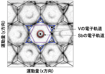
カゴメ格子は、三角形が頂点を共有して周期的に配列した結晶対称性を有 します。この特殊な幾何学的対称性は、電子の強いフラストレーションの 原因となり、様々な量子現象を引き起こすことが理論的に予言されていま した。なかでも CsV3Sb5 は、カゴメ格子では珍しい、超伝導を示すこと が2020 年に発見され、特殊な結晶対称性の下でなぜ超伝導が発現するか、 興味が持たれていました。本研究で「V 電子と Sb 電子の協調関係という 新たな効果」が超伝導発現の重要なカギであることが示されました。

光電子運動量顕微鏡で明らかにしたグラファイト 原子1 層のステップ構造

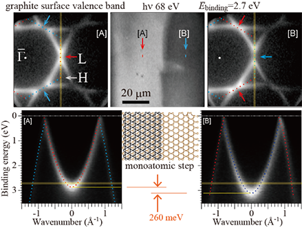
グラファイトの結晶は炭素のハチの巣状の原子層が積み重なる構造を持ち ます。表面を層に平行にへき開切断すると、奇数層目と偶数層目とで、そ れぞれ鏡面に映した関係の 3 回対称の構造が表面に現れます。しかし、従 来の測定では両者の表面構造を合わせて観測していたため、6 回対称の データが得られていました。そのため、これまでグラファイトの電子状態 は6 回対称であることが「常識」とされてきました。本研究では光電子運 動量顕微鏡を活用し、今まで気付かれていなかったグラファイトへき開最 表面の奇数層目と偶数層目の電子状態の違いを発見し、原子 1 層のステッ プの可視化に成功しました。

光電子運動量顕微鏡:マイクロメートルの機能性 材料の電子状態を空間・運動量分解能:50nm・0.01 Å-1 で可視化

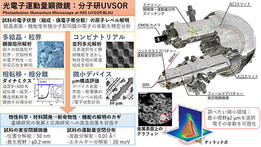
光電子分光は、X 線を試料表面に照射し飛び出す光電子を計測し、試料の 組成や電子物性を解明する分析法です。今回、UVSOR に最新の光電子分 光測定装置 Momentum Microscope を導入しました。不均一試料の 微小部分を拡大して観察できる顕微(microscope)機能と試料の物性を 決定づける電子のふるまい(運動量 momentum)を可視化する機能を1 つの装置で同時に実現します。本装置は主要な軽元素・遷移元素の内殻準 位を励起できる軟 X 線ビームラインに設置され、位置分解能 50 nm 、運 動量分解能 0.01 Å-1 、エネルギー分解能 20 meV を達成、試料を 9 K まで冷却でき、制限視野2 μm からの価電子帯分散やフェルミ面を測定す ることができます。本装置で初めて可能になる運動量分解顕微光電子分光 法は、表面・薄膜・配向分子・化合物結晶試料の原子レベルでの電子状態 解析を通じ、ナノ材料科学・量子デバイス工学を展開するうえで重要な手 法となります。

New Book released!

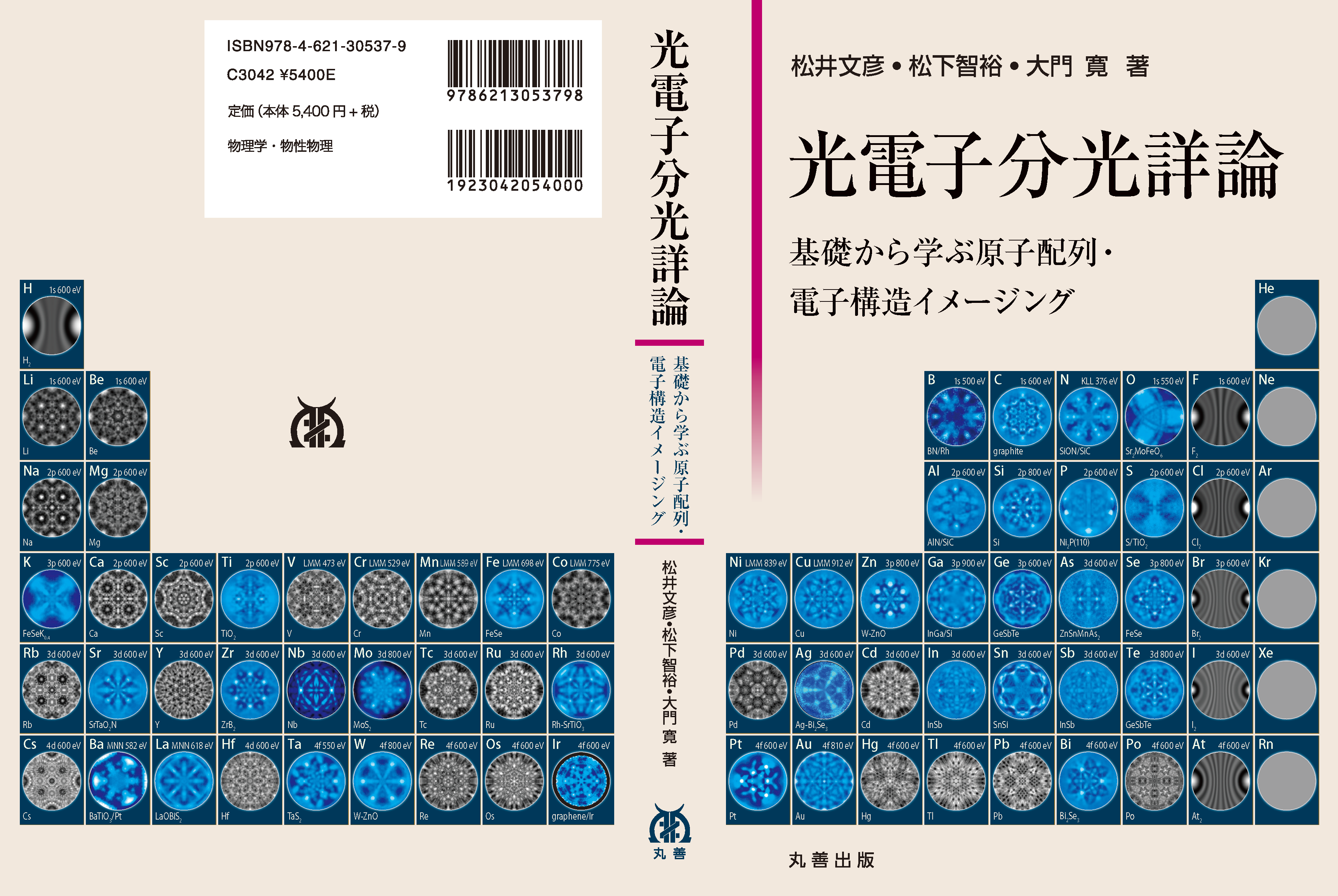
Textbook for photoelectron spectroscopy (in Japanese).
「光電子分光詳論」
authors: Fumihiko Matsui, Tomohiro Matsushita, and Hiroshi Daimon
ISBN: 978-4-621-30537-9 (Maruzen)
Photoelectron spectroscopy is widely used as a tool for investigating atomic structures and electronic states and clarifying physical properties, and is the basis for understanding physical physics and developing new materials. Advances in vacuum technology and the development of new light sources such as synchrotron radiation have made photoelectron spectroscopy an increasingly important method in the rapidly advancing materials science. On the other hand, in analysis, data that could not be seen until now is too visible now, so it is necessary to take a good look at the data itself. For that, it is necessary to return to the basics.
This book is a detailed commentary aimed at helping young researchers and students who begin to use photoelectron spectroscopy in earnest to learn the basics of photoelectron spectroscopy and analyze it by themselves. The two-dimensional anglar distribution woven by photoelectrons is very beautiful. This book is a guide to knowing and utilizing its beauty.

LINKS

ACCESS

〒444-8585
愛知県岡崎市明大寺町字西郷中38番地
38 Nishigo-Naka, Myodaiji, Okazaki 444-8585, Japan

アクセス情報
Access Information
自然科学研究機構 分子科学研究所 明大寺キャンパス 極端紫外光実験棟 Bld.9 (MAP)
National Institutes of Natural Sciences Institute for Molecular Science Myodaiji Campus UVSOR Bld.9
分子科学研究所 明大寺キャンパスまでのアクセスはこちら
Click here(https://www.ims.ac.jp/en/about/campus/access.html) for the access to Myodaiji Campus
極端紫外光研究施設(UVSOR棟) 301号室 TEL: 0564-55-7201
UVSOR Synchrotron Facility Building Room 301